Anatomy Of Musckes Sndctendons - Human Body Joint Anatomy Posters Muscles Tendons Bones Educational Chart Set Art Posters Art / The quadriceps are a collection of 4 muscles on the front of the thigh and are responsible for straightening the knee by bringing a bent knee to a straightened position.
bymamaheckman•
0
Anatomy Of Musckes Sndctendons - Human Body Joint Anatomy Posters Muscles Tendons Bones Educational Chart Set Art Posters Art / The quadriceps are a collection of 4 muscles on the front of the thigh and are responsible for straightening the knee by bringing a bent knee to a straightened position.. When the muscle contracts, the tendons are pulled, and the bone is moved. We did not find results for: Every skeletal muscle has three main parts: Related posts of anatomy of the foot muscles and tendons structure of anatomy leg and foot. A tendon connects the muscle to the bone.
The quadriceps are a collection of 4 muscles on the front of the thigh and are responsible for straightening the knee by bringing a bent knee to a straightened position. Extrinsic and intrinsic.the back functions are many, such as to house and protect the spinal cord, hold the body and head upright, and adjust the movements of the upper and lower limbs. By contracting, they also aid the venous return of blood to the heart and with age, these components of the musculoskeletal system progressively degenerate, which contributes to frailty. All together they help hold your upper arm in place in the shoulder. On the other hand, the insertion is where a tendon attaches that muscle to the *more* movable bone.
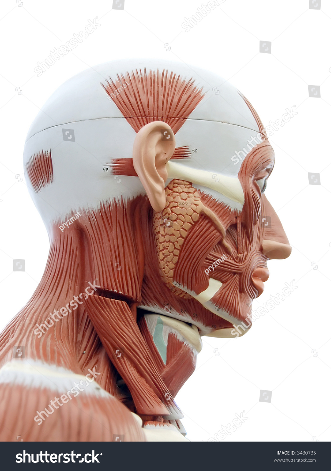
Human Anatomy Structure Head Muscles Tendons Stock Photo Edit Now 3430735 from image.shutterstock.com The quadriceps are a collection of 4 muscles on the front of the thigh and are responsible for straightening the knee by bringing a bent knee to a straightened position. The peroneal tendons run down together behind the outer side of the ankle and then split before attaching to different parts of the foot. Wrist anatomy is the study of the bones, ligaments and other structures in the wrist. On the other hand, the insertion is where a tendon attaches that muscle to the *more* movable bone. Winslow 2015 this essential companion book to the bestselling classic human anatomy provides artists and art students with a deeper understanding of human anatomy and different types of motion, The back is the body region between the neck and the gluteal regions. Originates from the upper part of the fibula, passes underneath the foot and attaches by the medial foot arch peroneus brevis: More specifically, this beautifully illustrated anatomy chart includes neck and shoulders, multiple views of the back and spine, and frontal views of each muscular extremity of the human body.
Each of them aids in a specific motion of your shoulder.
Schau dir angebote von muscle anatomy auf ebay an. *the origin, insertion, and belly.* a muscle's origin is where a tendon attaches it to the *less* movable bone. Bones in shoulder, ligaments of the shoulder joint, parts of the shoulder joint, shoulder anatomy, shoulder joints and muscles, shoulder structure anatomy, shoulder tendon anatomy, shoulder tendons ligaments, human muscles, bones in shoulder, ligaments of the shoulder joint, parts of. The wrist joint is a complex joint which connects the forearm to the hand, allowing a wide range of movement. A tendon connects the muscle to the bone. Über 7 millionen englischsprachige bücher. When the muscle contracts, the tendons are pulled, and the bone is moved. See tendons muscles foot lower leg anatomy stock video clips. Anatomy ankle anatomy ankle + ligament + tendon the foot anatomy human ankle anatomy 3d leg muscle lower leg anatomy leg articulation peroneal ankle muscles foot. The legs are the lower limbs of the human body that provide support and stability in addition to allowing movement. Tendons connect the knee bones to the leg muscles that move the knee. Originates from the upper part of the fibula, passes underneath the foot and attaches by the medial foot arch peroneus brevis: There are two main muscle groups around the knee:
Anatomy ankle anatomy ankle + ligament + tendon the foot anatomy human ankle anatomy 3d leg muscle lower leg anatomy leg articulation peroneal ankle muscles foot. 17 photos of the diagram of shoulder muscles and tendons. The legs include the upper leg, knee, lower leg, ankle, and. The peroneal muscles (peroneus longus and peroneus brevis), on the outside edge of the ankle and foot. There are two main muscle groups around the knee:
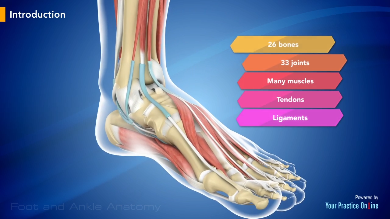
Foot And Ankle Anatomy Video Foot Ankle from www.ypo.education The quadriceps muscles provide strength and power with knee extension (straightening). The wrist joint is a complex joint which connects the forearm to the hand, allowing a wide range of movement. Maybe you would like to learn more about one of these? The peroneal tendons run down together behind the outer side of the ankle and then split before attaching to different parts of the foot. We did not find results for: Maybe you would like to learn more about one of these? By contracting, they also aid the venous return of blood to the heart and with age, these components of the musculoskeletal system progressively degenerate, which contributes to frailty. Lesson on the anatomy of the forearm:
Originates from the lower part of the fibula and attaches to the outer side of the midfoot
The peroneal tendons run down together behind the outer side of the ankle and then split before attaching to different parts of the foot. Über 7 millionen englischsprachige bücher. Tendons connect the knee bones to the leg muscles that move the knee. Extrinsic and intrinsic.the back functions are many, such as to house and protect the spinal cord, hold the body and head upright, and adjust the movements of the upper and lower limbs. Schau dir angebote von muscle anatomy auf ebay an. Winslow 2015 this essential companion book to the bestselling classic human anatomy provides artists and art students with a deeper understanding of human anatomy and different types of motion, The quadriceps and the hamstrings. However, it is susceptible to injury, especially from repetitive strain. More specifically, this beautifully illustrated anatomy chart includes neck and shoulders, multiple views of the back and spine, and frontal views of each muscular extremity of the human body. The fleshy, thick part of the muscle is called its belly. Originates from the upper part of the fibula, passes underneath the foot and attaches by the medial foot arch peroneus brevis: Maybe you would like to learn more about one of these? The majority of muscles in the leg are considered long muscles, in that they stretch great distances.
Maybe you would like to learn more about one of these? Check spelling or type a new query. The shoulder is not a single joint, but a complex arrangement of bones, ligaments, muscles, and tendons that is better called the shoulder girdle. Structure of anatomy leg and foot 6 photos of the structure of anatomy leg and foot leg foot anatomy, leg foot bones, leg foot cramps, leg foot cramps at night, leg foot massage, leg foot numbness, leg foot pain, leg foot tattoos, foot, leg foot anatomy, leg foot bones, leg foot cramps, leg foot. The primary function of the shoulder girdle is to give strength and range of motion to the arm.
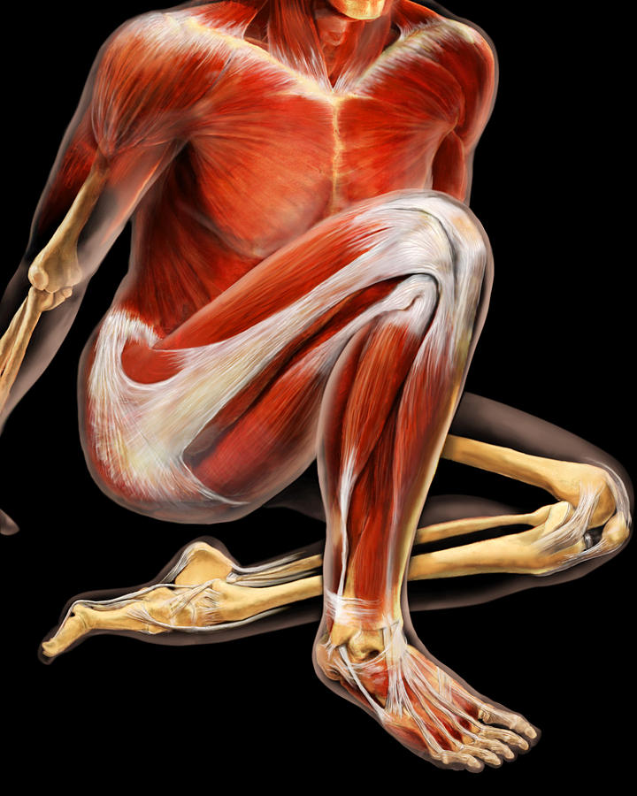
Muscles And Tendons Photograph By Anatomical Travelogue from images.fineartamerica.com Together, these muscles straighten your knee, stabilize your knee joint, assist in flexing your hip (drawing your knee towards your chest), and help absorb force when you land after jumping or leaping. Every skeletal muscle has three main parts: There are two main muscle groups around the knee: Maybe you would like to learn more about one of these? This muscular system chart shows in detail the deep layers of muscle on the back side of your body. Major muscles of the ankle. The quadriceps and the hamstrings. The fleshy, thick part of the muscle is called its belly.
It comprises the vertebral column (spine) and two compartments of back muscles;
Schau dir angebote von muscle anatomy auf ebay an. The quadriceps and the hamstrings. More specifically, this beautifully illustrated anatomy chart includes neck and shoulders, multiple views of the back and spine, and frontal views of each muscular extremity of the human body. When the muscle contracts, the tendons are pulled, and the bone is moved. Structure of anatomy leg and foot 6 photos of the structure of anatomy leg and foot leg foot anatomy, leg foot bones, leg foot cramps, leg foot cramps at night, leg foot massage, leg foot numbness, leg foot pain, leg foot tattoos, foot, leg foot anatomy, leg foot bones, leg foot cramps, leg foot. The fleshy, thick part of the muscle is called its belly. Related posts of anatomy of the foot muscles and tendons structure of anatomy leg and foot. Anatomy of musckes sndctendons / my english pages online: The quad muscles— which form the meaty mass on the front of your thighs — are among your strongest muscle groups, and play a critical role in athletic activities. Maybe you would like to learn more about one of these? The legs are the lower limbs of the human body that provide support and stability in addition to allowing movement. However, it is susceptible to injury, especially from repetitive strain. This is lesson 1 on the anatomy of the forearm.